

- Specification
- Downloads
- Reviews
$8,280.00
ADD TO CART

SKU: i357-20
References
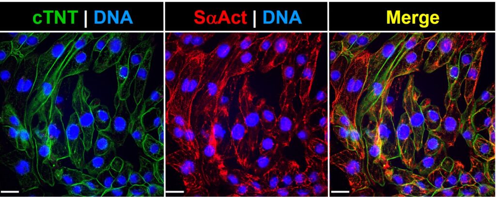
\n \nBrands:Cell Applications
Model Number:
Place of Origin:
Warranty:
Safety Performance:
Protection Class:
Sterile Status:
Reconstitution:
Shipping:
Battery:
Battery Service Life:
Component:
Capacity:
Concentration:
Dimension [D×W×H]:
Diameter:
Length:
Width:
High:
Volume:
Volume Range:
Speed:
Speed Range:
Max.Speed:
Max. RCF:
Temperature Range:
Operating Temperature Range:
Permissible Ambient Temperature :
Permissible Relative Humidity:
Input voltage:
Power Requirements:
Voltage/Frequency:
Sensitivity:
Specificity:
Precision:
Speed Accuracy:
Volume Accuracy:
Particle Size:
Pore Size:
Binding Capacity:
Calculated Molecular Weight:
Observed Molecular Weight:
Purity:
Shelf Life:
Storage:
Random Error:
Acceleration/Braking time:
Aspiration Speed:
Calculated Molecular Weight:
Dilution:
Driving Motor DC motor:
Em:
Ex:
Endotoxin:
Flow rate [liquid]:
Application:
Detection Target:
Detection Type:
Detection Range:
Research Areas:
Species Reactivity:
Reactivity:
Target Synonym:
Test Principle:
Quantitative/Qualitative :
Result Type:
Materials :
Assay Time:
Autoclave component:
Available Rotor Adapters:
Binding Capacity:
Bio-activity:
Bottom Shape:
Bottom Type:
Buffer:
Cap Color:
Clonality:
Conjugation:
Coupled Ligand:
Cover Type:
Detection Instrument :
Dispensing Speed:
Display:
Form:
Formulation:
Graduation:
Growth Area: